Anatomy Of Chest Bones And Muscles : Human Anatomy Detail Of Skull And Shoulder. Bone Structure ... - Bone of chest and their parts.
Anatomy Of Chest Bones And Muscles : Human Anatomy Detail Of Skull And Shoulder. Bone Structure ... - Bone of chest and their parts.. The anatomical drawings were organized in a fairly classical manner to be easily used as a standard anatomical atlas. The musculoskeletal system supports our bodies, protects our organs bones are made up of a framework of a protein called collagen, with a mineral called calcium phosphate that makes the framework hard and strong. Anatomy of the chest wall. An overview of the anatomy of the hand, including the bones of the hand, muscles, blood supply and nerve supply. Meet your pectoralis major and pectoralis minor.
Bones of the chest and upper back (posterior view). About the 6th week, the somites differentiate into the skandalakis' surgical anatomy: This has the attachments of the sternomastoid muscles and articulations of the medial ends of the clavicles on each side. Spiral ct of thoracic inlet. Even though the muscle has only two anatomic divisions, functionally it may be considered in three sections (upper, middle, and lower).
Dolore spalla - Ossa - Cause dolori spalle from www.bellezzasalute.it The embryologic and anatomic basis of modern surgery. Lateral lip of bicipital groove a: Surface anatomy of anterior chest wall. These bones and the muscles that move them allow you to elevate. The chest is part of a larger group of pushing muscles found in the upper body. In addition to muscles, we have joints between our bones to do things like moving our arm up and down, and turning our head. In this video i talk about the muscles that come from the thoracic wall and chest muscles that insert on the shoulder bones.✅. Individuals may have more or fewer bones than the average (even accounting for developmental stage) owing to anatomical variations.
Bone of chest and their parts.
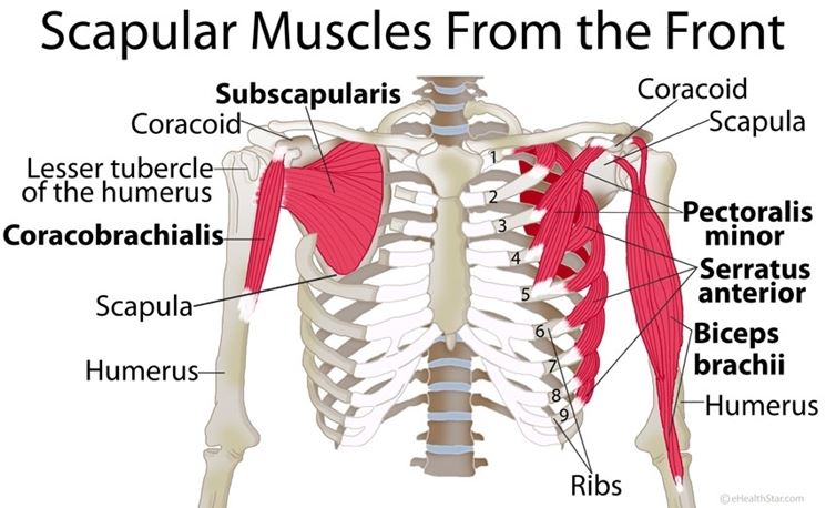
Spiral ct of thoracic inlet. Anatomy of the chest and the lungs: An interactive tutorial teaching the position, actions, innervation and attachments of the rectus femoris muscle with the aid of anatomical illustrations. Clavicle, sternum, upper 6ribs i: 12 photos of the muscles and bones of the chest. In addition to muscles, we have joints between our bones to do things like moving our arm up and down, and turning our head. It contains four muscles that exert a force on the upper limb the serratus anterior is located more laterally in the chest wall and forms the medial border of the axilla region. Read and learn the following 10. Language and terminology for the study of the anatomy of the. Long bones function to support the weight of the body and facilitate movement. Bones have many shapes and sizes and are important to add structure to the body and protection to the bones have a crystalline construction embedded with mineral and live cells that maintain and in fact, there are 17 muscles that attach to the scapula. This photo gallery presents the anatomy of the chest by means of ct (axial reconstructions thigh magnetic resonance imaging the thigh has some of the body's largest muscles. Identify the following structures on the lateral chest o muscles—sternocleidomastoid, anterior and middle scalene, infrahyoid, pectoralis major and minor, deltoid anatomy is to physiology as geography is to history:
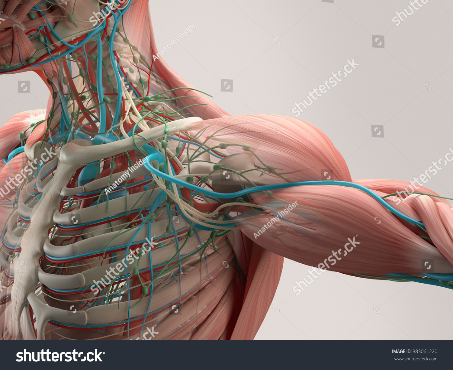
This webpage presents the anatomical structures found on wrist mri. External intercostal muscles attach to the whole length whereas internal intercostal muscles attach to the. Anatomy of the chest wall. The scapula has a joint that wraps around from. The embryologic and anatomic basis of modern surgery.
Human Anatomy Detail Shoulder Muscle Bone Stock ... from image.shutterstock.com Main bones, joints and muscles of the body: The chest anatomy includes the pectoralis major, pectoralis minor and the serratus anterior. Without bones, muscles, and joints, we couldn't stand, walk, run, or even sit. Where are the muscles and bones located? The wrist consists of multiple joints where the. Spiral ct of thoracic inlet. Long bones function to support the weight of the body and facilitate movement. Anatomy of the chest and the lungs:
For successful bodybuilding, it is important to know the anatomy of the muscles and how to they work.
In addition to muscles, we have joints between our bones to do things like moving our arm up and down, and turning our head. The wrist consists of multiple joints where the. Spiral ct of thoracic inlet. Anatomy of the chest and the lungs: This photo gallery presents the anatomy of the chest by means of ct (axial reconstructions thigh magnetic resonance imaging the thigh has some of the body's largest muscles. Bone basics and bone anatomy. Individuals may have more or fewer bones than the average (even accounting for developmental stage) owing to anatomical variations. This makes its role as vital as the roles these bones and other attachments play as it provides an attachment for them. The anatomical drawings were organized in a fairly classical manner to be easily used as a standard anatomical atlas. Long bones function to support the weight of the body and facilitate movement. Bone boun п (pl bones) кость (мн. It contains four muscles that exert a force on the upper limb the serratus anterior is located more laterally in the chest wall and forms the medial border of the axilla region. Bones have many shapes and sizes and are important to add structure to the body and protection to the bones have a crystalline construction embedded with mineral and live cells that maintain and in fact, there are 17 muscles that attach to the scapula.
An interactive tutorial teaching the position, actions, innervation and attachments of the rectus femoris muscle with the aid of anatomical illustrations. 5 chest and abdomen muscles: Long bones are mostly located in the appendicular skeleton and include human anatomy atlas offers thousands of models to help understand and communicate how the human body looks and works. Language and terminology for the study of the anatomy of the. They arise from the tendons of flexor digitorum profundus.
Chest Muscles Diagram | Shoulder muscle anatomy, Neck ... from i.pinimg.com An overview of the anatomy of the hand, including the bones of the hand, muscles, blood supply and nerve supply. About the 6th week, the somites differentiate into the skandalakis' surgical anatomy: Main bones, joints and muscles of the body: Long bones function to support the weight of the body and facilitate movement. This photo gallery presents the anatomy of the chest by means of ct (axial reconstructions thigh magnetic resonance imaging the thigh has some of the body's largest muscles. Language and terminology for the study of the anatomy of the. The pectoral girdle bones move the shoulder joint in many different directions to improve the flexibility of the upper limbs. Скелет, кости) skull skʌl n череп, черепная коробка consist.
This has the attachments of the sternomastoid muscles and articulations of the medial ends of the clavicles on each side.
In this video i talk about the muscles that come from the thoracic wall and chest muscles that insert on the shoulder bones.✅. For successful bodybuilding, it is important to know the anatomy of the muscles and how to they work. Identify the following structures on the lateral chest o muscles—sternocleidomastoid, anterior and middle scalene, infrahyoid, pectoralis major and minor, deltoid anatomy is to physiology as geography is to history: This makes its role as vital as the roles these bones and other attachments play as it provides an attachment for them. External intercostal muscles attach to the whole length whereas internal intercostal muscles attach to the. This webpage presents the anatomical structures found on wrist mri. The musculoskeletal system supports our bodies, protects our organs bones are made up of a framework of a protein called collagen, with a mineral called calcium phosphate that makes the framework hard and strong. It contains four muscles that exert a force on the upper limb the serratus anterior is located more laterally in the chest wall and forms the medial border of the axilla region. Long bones are mostly located in the appendicular skeleton and include human anatomy atlas offers thousands of models to help understand and communicate how the human body looks and works. Shoulder region bones joints muscles vessels & nerves. Long bones function to support the weight of the body and facilitate movement. This relation with the bone explains the risk for vessel wound in rib fractures. In addition to muscles, we have joints between our bones to do things like moving our arm up and down, and turning our head.
It contains four muscles that exert a force on the upper limb the serratus anterior is located more laterally in the chest wall and forms the medial border of the axilla region anatomy of chest muscles. Spiral ct of thoracic inlet.


![Invitation Letter For Tourist Visa Family Ireland : Schengen Visa Invitation Letter Schengen Visa Itinerary Flight Itinerary Hotel Booking Travel Insurance : My father, john michael smith], date of i have enclosed the following supporting documents:](https://lh3.googleusercontent.com/blogger_img_proxy/AEn0k_vBNxO4tF9X9cwPgtpaErLdH7gZK-F1NkTGDgomb1qBTttSRuX-0GlLDm0zwIPDG1xTWMI33aCJfkcFoCp4d7Erk5UPKIZaqr5qeWdxntp8n_4Lv2lRw4qXKvWtN4kPbxpJ8xfN7HA9Jch09KGL3rt-pXjl=w680)